

PT Wound Care: Assessment, Dressings and Dressing Changes

The patient is already positioned, the old dressing is half lifted, and someone is asking which foam to open. That is the moment PT wound care usually drifts off course. The product is the last decision, not the first.
A heel that has stayed open for weeks, a sacral injury that looks unchanged after two changes, or a shin wound under a wrap that was never checked for pulses all share the same miss. The team treated the cover and left the cause alone. Assessment, off-loading, and a clear stop-and-refer line decide whether tissue moves this week. The dressing only holds the conditions those decisions create.
If the chart has no baseline length, width, and depth, this visit cannot show change. If the limb is ischemic, compression can injure tissue before the next shift. Build the plan from the wound, the blood flow, and the pressure. Then match the cover to that plan.
Pre-Treatment Checks That Decide the Plan
A 68-year-old with type 2 diabetes arrives with a heel ulcer that has been open for three weeks. The first visit decides whether the next month is healing, a stall, or a referral that should have happened today. Chronic wounds affected approximately 10.5 million Medicare beneficiaries, about one in six, in an updated compendium available through PMC. That scale is why the baseline check outranks the dressing on the cart.
Classify the wound before anything else. Location, type, and depth come first, then a screen for infection, edema, diabetes control, pressure, and nutrition. The American Academy of Family Physicians lower-extremity sequence puts perfusion ahead of compression, using ABI or, when calcification is suspected, toe pressure or vascular testing. Debride devitalized tissue when it is clinically appropriate, keep the bed moist rather than macerated, treat the cause, and record dimensions for later comparison.
ABI is only as good as the vessel. It can read falsely acceptable in calcified arteries and in some heel wounds. Rest pain, gangrene, or visible tissue loss overrides a comfortable number. Do not wrap a limb because the printout looks fine while the foot is cold and the patient cannot sleep. If the ABI cannot be obtained or the story does not fit the number, stop and get vascular input before any wrap goes on.

What the first numbers lock in
An American Academy of Family Physicians review of lower-extremity wound care of an observational dataset involving 154,664 patients and 312,744 wounds reported a 70.8% overall healing rate when debridement was performed consistently at intervals of one week or less, with faster healing than less frequent or inconsistent treatment. Debride on a weekly or shorter interval, within the team's scope.
Note infection signs before the cover goes on. Record length, width, and depth before the first dressing so the next visit has a real comparison. Tie that record to prevention practice such as the wound care nursing diagnosis for pressure ulcers, so off-loading starts with the plan rather than after the skin fails again.
Baseline measurements and the plan, including the ABI result, the compression decision, and the next reassessment date.
Those two lines are the visit. Without them, the next clinician inherits a dressing instead of a trajectory.
Choosing a Dressing for the Wound You Have
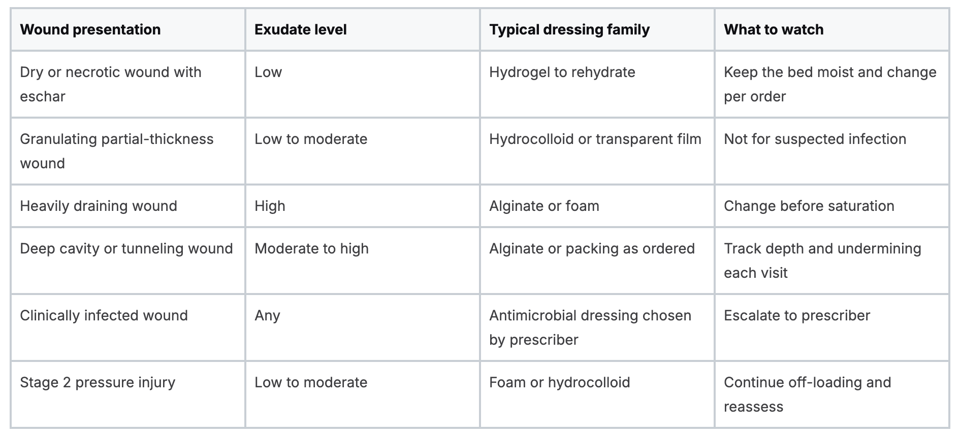
The order on the chart and the wound in front of you pick the dressing. Habit does not. Match exudate, depth, and infection risk first, then check whether the patient can wear that choice.

Filters that override the table
Three practical limits change a correct family into a failed one. Ask how often the patient or caregiver can change the dressing, whether the patient is up and walking, and whether the cover stays put under compression.
A patient with a shin wound who lives alone and cannot reach a daily change should get a longer-wear foam, not a dressing that needs attention every morning. If the same limb needs compression for venous disease, the compression tips for venous ulcer care from Vein and Wound Institute are a useful check on how the wrap and the dressing have to work together. A bulky island that buckles under the bandage is the wrong product even when the exudate column looks right.
Never leave a hydrocolloid on a wound you suspect is infected.
Hydrocolloid and transparent film belong on clean, low-drainage beds. An antimicrobial dressing is a prescriber choice, and it does not replace a same-day call when redness is spreading. For clinicians building that selection skill, Wound CareDressings 101 covers basic through advanced techniques for selecting and applying dressings that support outcomes. The course does not replace the order in the chart.
How to Perform a Dressing Change Step by Step
A clean change protects the wound edge and gives you a measurement you can trust. Rushing the removal, then grabbing a forgotten supply, is how the field gets contaminated and the skin gets torn.
The short flow below groups the work. The numbered sequence after it is what you do at the bedside.
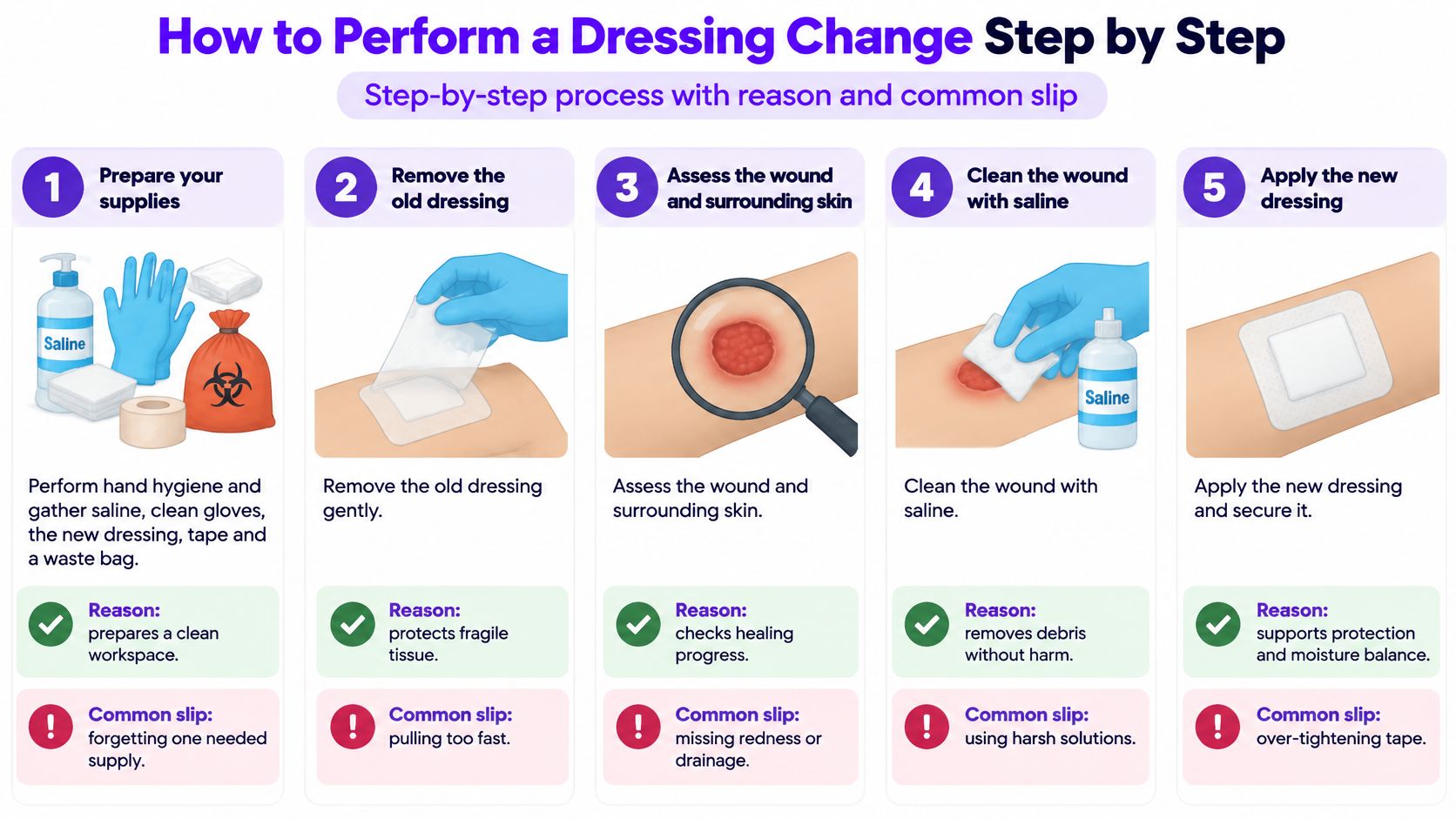
- Set the field first. Perform hand hygiene and gather saline, clean gloves, the new dressing, tape, and a waste bag. Reaching back into a drawer mid-procedure drags the clean field into the dirty one. If blood or body fluid exposure is part of the job, the OSHA bloodborne pathogens course is the companion standard for how that exposure is handled, separate from the dressing steps themselves.
- Position and explain. Turn the patient so you can see the whole wound, and say what you are about to do. A patient who knows the next move tolerates the pull better and is less likely to jerk the limb.
- Remove slowly. Wet adhesive with saline so skin and wound edges are not stripped, then check for residue. Yanking a dry border is the slip that turns a partial-thickness edge into a larger injury.
- Inspect before you clean. Measure the wound and look at periwound skin, drainage color, and odor. Skipping this because the clock is short means the note describes the old dressing, not the tissue.
- Cleanse from the center out. Use saline or a pH-balanced cleanser around 5.5. The American Academy of Family Physicians pressure-injury guidance notes that standard hospital soaps may have a pH of 9.5 to 10.5, while a balanced cleanser may sit near 5.5, and it advises against highly alkaline soaps. Scrubbing outward-in drags periwound bacteria into the bed.
- Protect the border. Pat the periwound dry and apply a moisture barrier where incontinence is present. Leaving the skin wet under the new cover is how maceration shows up at the next visit. The same guidance says prevention starts as soon as risk is known: for an immobile patient admitted after a stroke, staff assess risk, inspect skin, support nutrition, reposition, and choose a support surface before breakdown is visible.
- Apply without tension. Seat the dressing with margins on intact skin, with no wrinkles and no tight pull. Wrinkles become pressure points. Tape that blanches the edge is too tight.
- Label the change. Write the date and initials, and set the next change date where the next person will see it. An unlabeled border invites an early peel "just to check."
- Document what you saw. Record the change, the measurements, and the drainage. A note that only says "dressing changed" cannot show healing or a stall.
A sound change leaves a moist bed, drainage that is controlled, periwound skin that is not white and soggy, and a patient who is comfortable enough to move as planned.
Off-Loading and Debridement Timing
The dressing cannot heal a wound that is still being sat on, stood on, or pinched by a device. Off-loading is the treatment. The cover is the protection around it.
- Find the pressure. Check heels, sacrum, trochanters, and skin under splints, braces, and seating. A new redness ring under a boot strap is a device injury, not a dressing failure.
- Set a repositioning schedule. For patients in bed, that is typically every two hours, or whatever the plan orders, adjusted for the support surface and what the patient can tolerate.
- Float heel wounds. Use a pillow or an off-loading boot so the calf carries the weight and the heel does not touch the mattress.
- Recheck the surface. Confirm the pressure-redistributing mattress or cushion at every visit. A deflated cell is not off-loading.
- Add movement. Build sit-to-stand practice, supported transfers, and a caregiver routine for repositioning into the same plan as the dressing.
Say a patient with a plantar heel ulcer spends most of the day in a recliner. The visit sets a pressure-relief schedule, floats the heel, and the next visit includes a look at that chair cushion.
Remove devitalized tissue when it is clinically appropriate and inside the treating team's scope. Schedule the interval, write it down, and coordinate with the nurse or surgeon who performs sharp debridement rather than assuming someone else already did it. Electrical stimulation has supportive guideline-level evidence as an adjunct. It complements pressure redistribution, tissue management, infection control, nutrition, and mobility work. It does not replace them.
Monitoring for Infection and Stalled Healing

A single wound photo records a moment. Healing shows up in the trend: area, depth, undermining, drainage, and pain measured the same way at each visit. The multidisciplinary pressure-injury approach summarized in PMC depends on that discipline. Stage and measure consistently, identify the drivers of pressure, off-load, manage the bed and support surface, restore mobility, then compare serial numbers rather than a fresh impression. PT earns a place in that loop because mobility work lowers immobility-related risk, and positioning and exercise remain part of care after the injury appears.
A sacral injury measured at baseline and then at each weekly review makes the point. If the area stays flat across three measurements while the patient remains on the same mattress, reassess off-loading and the surface before changing the dressing. Swapping foam when shear and time in the chair are unchanged treats the supply closet, not the wound.
Where the decision sits
The escalate column is the clinical decision. New undermining, tunneling, or exposed bone suggests deeper infection and calls for referral, not another week with the same cover. Early local signs include spreading redness beyond the wound edge, warmth, induration, purulent or foul drainage, and pain that is new or increasing. I treat new or rising pain as a legitimate trigger even when the wound looks quiet; the table is not a substitute for that exam.
Same-day calls
Fever, chills, or confusion warrant a same-day call to the prescriber. Those findings do not belong in the next team huddle. A chronic wound that has stalled also needs a structured review of the reasons it is not closing. Why Won't This Wound Heal addresses management of non-healing wounds and how to work with patients through that review. Local protocol still sets the bedside plan.
Common misses: treating the dressing while pressure, shear, or immobility stay unchanged, and using a modality as a substitute for off-loading and wound-bed preparation. Device-related pressure gets the same reassessment as the mattress, including straps, braces, and seating edges.
Staging, Documentation and Patient Teaching
Stage by the deepest tissue involved, not by how dramatic the wound looks. The Wolters Kluwer pressure injury pocket card states the mechanics plainly. Stage 1 is intact skin with localized, non-blanchable erythema. Stage 2 is partial-thickness loss with exposed dermis and a pink or red, moist bed. Stage 3 is full-thickness loss in which adipose tissue may be visible, and Stage 4 reaches deeper tissue. If slough or eschar completely obscures the base, the injury is unstageable.
A shallow ruptured serum-filled blister can look severe. Document the actual depth. It is not a Stage 3 wound because it startled someone.

Use the same checklist every visit so the record stays audit-ready under facility policy.
- Date and time: when the assessment happened.
- Location and size: site, plus length, width, and depth.
- Stage and drainage: deepest tissue and what is coming out.
- Border and cover: periwound condition and the dressing applied.
- Response and plan: tolerance, who was notified, and the next review date.
At home, the patient keeps skin clean and hydrated, uses a moisture barrier after incontinence, shifts on the team's schedule, and reports new redness, odor, fever, or a dressing that is soaked or loose. Before they leave, use a teach-back: "Show me what you will do if the dressing comes loose tonight." Confirm the answer. A nod is not teaching.
When PT Leads, When to Refer Out and Next Steps
Physical therapists earn their place in wound care through edema control, off-loading, exercise, mobility, positioning, and the handoff to self-management. Those interventions change healing and function, and they stay underused. A wound-care visit is a shared exam. Function is the PT contribution.
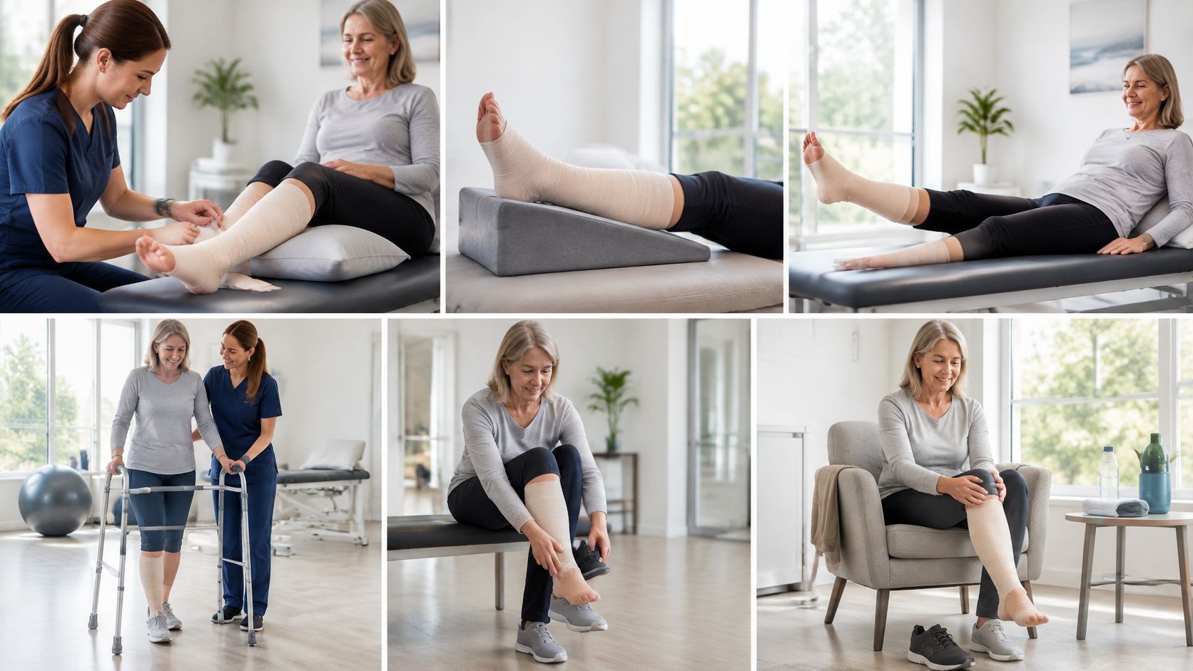
The Advances in Skin & Wound Care review of PT wound practice notes that these interventions have evidence support, yet therapists remain underused partly because of reimbursement structures and limited awareness of what they can do. Reimbursement rules, administrative burden, unclear eligibility, and state variation in whether PTs may perform sharp debridement all constrain practice. Proposed models do not consistently require coordination with surgeons and primary care. Adding techniques without an escalation path fragments care.
PT improves function. It does not replace medical wound assessment, vascular evaluation, infection management, or debridement. Stop and refer the same day when any of these show up.
A PT treating a venous leg ulcer confirms perfusion, applies compression only inside the allowed range, and coordinates changes with nursing. When drainage turns purulent and the patient spikes a fever, the visit stops, the note is written, and the referral goes out that day. Healing, function, cost, and patient selection in PT-managed chronic wounds are not yet well measured. Do not sell process convenience as proof of faster closure.
Before the next clinic day, confirm the facility ABI and compression rule, the dressing formulary and who orders changes, and a dressing log so the trend is visible. Course information is not meant to serve as a guideline for patient management. Optional structured training includes Wound Care Dressings 101 for selection and application, and Why Won't This Wound Heal for non-healing wounds. This week, confirm one protocol item: who on your unit is authorized to compress, debride, and escalate.
The catalog includes ANCC-accredited nursing continuing education with wound-care courses on dressing selection and non-healing wounds, plus state requirement guidance for clinicians who need renewal credit. The same catalog supports healthcare teams and school staff administrators who assign and track training. Review courses and set up the training your role requires at Premiere Education.
Latest Posts





25% off Sitewide→
Use Code: PCECYBERSALE2025→