

Large Body Habitus: Definition and Clinical Adaptations
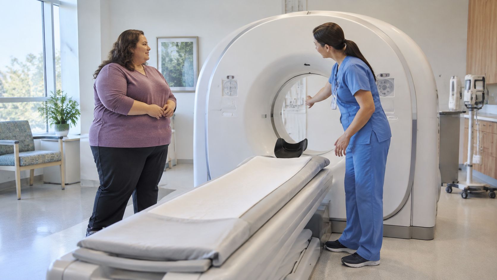
A booking comes in for a contrast CT. The referral lists height and weight, but no girth, no mobility notes, and no check of the scanner table. By the time the patient is on a stretcher in a gown that doesn't close, the team is solving problems that intake could've settled in five minutes.
That's the practical issue with large body habitus. The fix isn't working harder on the day. It's recording the right measures, matching them to equipment limits, choosing the right weight for each drug, and planning the transfer before transport starts.
What Large Body Habitus Means and Where BMI Falls Short
The referral lists 150 kg, height, and a contrast CT. No girth. No mobility notes. No dosing weight. The largest gown in the cupboard is too small and the stretcher is standard width. The team failed at sizing the job before the patient arrived.
Large body habitus describes overall body size and shape as it changes how care is delivered. It determines scanner fit, dose calculation, transfer staffing, and cuff accuracy. It functions as a planning term for the room and the roster.
What BMI Tells Clinicians, and What It Doesn't
The World Health Organization obesity and overweight fact sheet defines adult overweight as a body mass index of at least 25 kg/m² and obesity as a BMI of at least 30 kg/m². The same fact sheet reports that in 2024, approximately 2.6 billion adults aged 18 years and older, 45% of the global adult population, were overweight, including about 940 million adults living with obesity.
Global adult obesity prevalence had more than doubled between 1990 and 2024, reaching approximately 16% in 2024, according to the World Health Organization obesity and overweight fact sheet. Larger patients are a routine part of the list. Rooms, stock, and workflows either account for that volume or they scramble every shift.
BMI is built from weight and height only. It does not separate muscle from fat, locate where tissue sits, or describe mobility, strength, or tolerance for lying flat. Two patients can share a BMI and need different scanners, different cuffs, and different transfer plans.
BMI sketches the outline. Equipment and drug decisions require direct measurements.
The measures that drive planning are concrete. Teams need weight for load ratings, height for weight descriptors, waist or girth for cuffs and positioning, and maximum transverse body diameter for bore fit. Add mobility and transfer ability. With those five items at intake, the team books the right scanner, selects a dosing weight, and staffs the move correctly.
Imaging Equipment Limits and Scanning Constraints
Large body habitus creates capacity and geometry problems that start before image quality is even considered. The table rating decides if the patient can be lifted safely. The bore decides if the scan can happen at all. I recommend logging weight, maximum transverse diameter, and the exact scanner model at intake, so booking never relies on memory.
The review of imaging constraints in obesity reports industry-standard limits of approximately 450 lb (204 kg) and 70 cm aperture for CT, 350 lb (159 kg) and 60 cm for cylindrical-bore MRI, and 550 lb (249 kg) with a 55 cm maximum diameter for vertical-field open MRI. Those figures are starting points. The exact model in the department sets the actual limit.
Imaging Equipment Limits for Large Body Habitus

Why Diameter Beats Weight as a Fit Test
A 58 cm transverse diameter in a 60 cm bore leaves about 1 cm per side. Coils, padding, clothing, and arm position still need space. A table 15 to 18 cm thick reduces the usable vertical opening further, so a patient can pass the weight check and still not fit safely or comfortably.
That trade-off belongs at booking. A forced fit risks pressure injury, panic, aborted sequences, and a second appointment. Measuring diameter at intake takes a minute. It prevents transporting a patient to a scanner that was never going to work.
Radiography fails differently. Increased tissue thickness degrades penetration and contrast, so the radiographer adjusts exposure technique, positioning, grid use, and collimation. The practical example is an abdominal film where bowel gas detail washes out. Raising exposure without a plan improves penetration while it worsens scatter, so technique changes need to stay deliberate.
Ultrasound, especially abdominal ultrasound, is limited by depth. Sound weakens as it travels through more tissue, and standard presets may not show the liver edge, gallbladder, or pelvic structures clearly. Use a lower frequency transducer, switch on harmonics, reposition the patient to shorten the beam path, and book a longer slot. Name the fallback in advance too. If ultrasound cannot answer the question, CT or MRI becomes the booked next step, not an afterthought after a non-diagnostic scan.
Scheduling holds when three details are fixed before transport. The request records weight and maximum transverse diameter, a staff member phones to verify the exact scanner limits, and the notes name an alternative scanner or referral pathway.
Adjusting Medication Doses Beyond Total Body Weight
Bigger does not automatically mean a proportionally bigger dose. Fat and lean tissue handle drugs differently, and the wrong weight descriptor can overdose one drug while underdosing another in the same patient. Plan the descriptor at intake, alongside the scanner limit and the transfer step, so prescribing does not start from total body weight by default.
A pharmacokinetics review on dosing in obesity explains that adipose tissue has relatively low metabolic activity, so drug clearance often correlates more closely with lean body weight than total body weight. Using total body weight for maintenance dosing can therefore increase adverse-event risk.
Loading Doses and Maintenance Doses Behave Differently
Lipophilic drugs distribute into fat. For some loading doses, total body weight matters more because the drug must fill a larger volume of distribution to reach target concentration. Maintenance dosing follows clearance. Clearance usually tracks lean tissue, kidney function, and liver function more than fat mass.
Volume of distribution may increase, remain similar, or change disproportionately depending on the drug. Clearance can decrease, increase, or remain unchanged depending on the elimination pathway. The same patient may need total weight for one calculation, adjusted weight for a second drug, and ideal weight for a third. I recommend recording which descriptor applies to each charted drug before the patient reaches the scanner room.
A Worked 130 kg Example
A man 170 cm tall weighing 130 kg has an ideal body weight of about 66 kg. His excess weight is about 64 kg, and an adjusted weight using ideal plus 40% of the excess comes to about 92 kg. Those three numbers, 66 kg, 92 kg, and 130 kg, produce very different milligram doses for any weight-based prescription.
For a drug confined mainly to lean tissue, dosing from 130 kg could deliver close to double the exposure intended from ideal weight. For a lipophilic loading dose, dosing from 66 kg could leave the patient under-treated because a large share of drug has moved into fat. Charts should show the descriptor, the formula, and the arithmetic alongside the final dose.
Weight Descriptors for Dosing in Large Body Habitus

Aminoglycosides give a practical illustration. Many institutional protocols use adjusted body weight because these drugs distribute into extracellular fluid, which increases somewhat with larger body size but not in step with fat. Ideal weight alone can underdose, and total weight can push concentrations into toxic range. Levels and renal function then decide whether the estimate was right.
Practical rule: Choose the descriptor from drug-specific evidence, then let response, toxicity checks, and drug levels correct the estimate.
Renal and hepatic function still matter alongside any weight. A carefully chosen descriptor will not rescue a maintenance dose if kidney clearance has fallen or liver metabolism is impaired. Titration should use clinical response, toxicity surveillance, and therapeutic drug monitoring when it is available.
If the patient is not responding or shows toxicity, change the dose based on that evidence, not on loyalty to the original calculation. Documentation prevents the next shift from recalculating blindly. The prescription note should state measured weight, descriptor used, and reason, for example adjusted weight for an aminoglycoside per local protocol.
Communicating With Dignity and Protecting Privacy
Measurements only help if the patient agrees to them. A patient who feels exposed may refuse the scale, estimate a weight from months ago, or cancel the scan. Phrasing decides whether girth and diameter get recorded accurately or become the point where care stalls.
Explain the reason before touching the tape measure. A useful line is, "The scanner opening is a fixed size, so a measurement around the widest point tells the team which scanner can take the images safely." That framing puts the constraint on the equipment, not on the patient's body, and it gives the measurement a purpose the patient can accept.
Phrases That Keep Cooperation
- Ask permission first: "Is it okay to measure and to talk through weight for the medication calculation?"
- Name the equipment limit: "That table has a load rating, so the team checks it before any transfer."
- Offer control where possible: "Would a closed room and one staff member be better for weighing?"
- Avoid blame language: Replace "too big for the scanner" with "this scanner's opening isn't wide enough, so the booking goes to the larger unit."
Privacy needs the same advance planning as the transfer step. Weighing belongs in a closed room on a scale rated for the patient, with the reading visible only to staff who need it. Keep weight off open charts, whiteboards, or waiting-room screens. Have a gown that fits in the room before the patient is asked to change, not fetched while the patient waits uncovered. Confirm the scanner assignment and the weight used for dose checks at intake, so the bedside conversation stays short.
In a shared bay, five staff gather around a stretcher to discuss whether the table will hold, and the patient declines the scan. The clinical concern is legitimate, but the audience makes it humiliating. Assign a single named lead, move to a private space, and agree the transfer plan before extra staff enter. Fewer voices, closed curtains, and a gown that closes turn a refusal into a completed examination more often than persuasion does.
Staff who want concrete language and policy examples can use guidance on preventing weight discrimination in healthcare settings alongside local procedures. The aim is to remove the moments that make patients hide information the team needs for safe dosing and imaging.
Documentation should stay factual. Record the measurement, the reason it was taken, and the patient's own account of mobility, transfer needs, and past experiences with scanners or sedation. Write "transfers with a hoist and two staff" rather than a judgment about ability. The next team gets instructions it can act on, and the patient is not reduced to a label in the notes.
Preparing a Large-Bodied Patient and Building the Clinic Protocol
The booking looks straightforward until transfer time exposes what intake missed. A scanner is free, the patient has arrived, and then the table rating, the doorway width, or the lack of a hoist stops the list. Failures cluster at the transfer step, and that is why the check of scanner limit, dose weight, and transfer plan has to happen before the patient travels. The Association for Radiologic Imaging Nursing bariatric patient safety guidance recommends advance assessment of height, weight, BMI, girth, comorbidities, prior sedation history, and equipment needs. It also identifies higher-capacity wheelchairs and stretchers, larger gowns, appropriately sized monitoring devices, transfer aids, pressure-point padding, wider doorways, and additional staff for safe transfers.
The six-step overview below gives the sequence at a glance.
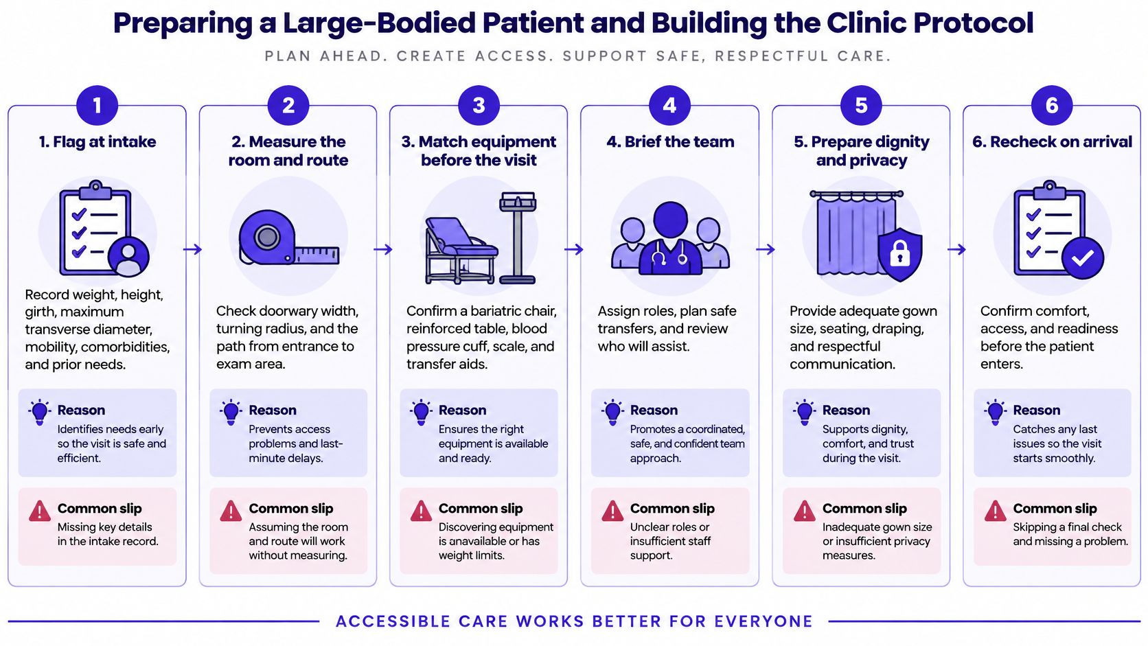
The Six Steps Before the Patient Arrives
- Flag at intake: Record weight, height, girth, maximum transverse diameter, mobility, comorbidities, and prior sedation history at booking. Every later decision, scanner choice, cuff size, and staffing, depends on those numbers. The common slip is accepting a self-reported weight that's months old.
- Verify equipment: Confirm scanner and table limits, then check that a bariatric stretcher or wheelchair, larger gowns, and a correctly sized blood pressure cuff are physically available. The common slip is assuming that a bariatric waiting chair means the scanner table is also bariatric rated.
- Plan the transfer: Set staff numbers, choose transfer aids such as a slide sheet or hoist, add pressure-point padding, and check doorway width and turning space on the route. Planning the transfer on the day, with the patient waiting, is how staff injuries and skin damage happen.
- Confirm the dosing weight: Agree the weight descriptor for each prescribed drug and document the calculation in the chart. Defaulting to total body weight for every drug is the slip this step exists to prevent.
- Set monitoring and access: Anticipate longer needles or ultrasound-guided IV access, an alternative blood pressure method if a standard cuff won't fit or read reliably, and sedation and airway planning that accounts for positioning. A ramped position and extra hands for mask ventilation should be decided before sedation starts, not during it.
- Escalate or refer early: If any limit is exceeded, move the booking to suitable equipment or another site before transport. Referral is part of safe care when the equipment can't take the patient.
Turning Steps Into a Written Protocol
A checklist that lives in one nurse's memory breaks on annual leave. The clinic protocol needs one named owner who keeps it current. That owner maintains the equipment list, checks expiry and load-rating labels, and reviews the document after any incident, near miss, or aborted scan. I recommend placing ownership with the lead who already signs off scanner bookings, so updates reach the rota without a second handover.
The equipment list should be specific enough to use at 2 a.m. For each scanner and bed, it states the weight limit, the aperture or bore size, and where the higher-capacity stretcher, hoist, large cuffs, and gowns are stored. "Bariatric equipment available" isn't a usable entry. A location and a rating are.
A referral pathway belongs on the same page. It names the alternative site, the phone number, and what measurements that site needs before accepting the booking. Staff shouldn't have to negotiate from scratch while a patient sits in a corridor.
The dosing reference needs the same treatment. For each high-risk drug class the clinic uses often, such as aminoglycosides, anticoagulants, and sedative agents, the reference names the weight descriptor, the formula, and the monitoring that follows. Pharmacy should approve that page, and prescribers should be able to open it from the prescribing screen. Background reading on best practices for providing inclusive healthcare can support the service standards around the protocol, but local equipment ratings still govern daily decisions.
Suitable furniture and equipment are part of clinical care, not an optional extra added when a patient asks.
Rechecking is built into the protocol, not left to goodwill. Scanner limits get verified whenever equipment changes, and the list gets reviewed after any incident. During a refurbishment, for example, a CT table gets replaced with a different rating. If the protocol still carries the old rating, the next booking decision is wrong even though every staff member followed the document.
Escalation triggers close the loop. Staff escalate when a weight or diameter limit is exceeded, when IV access fails after the agreed number of attempts, when blood pressure can't be measured reliably, or when a transfer can't be done safely with available staff and aids. Naming those triggers in advance gives a junior nurse permission to stop and call for help before an unsafe move, rather than apologizing after one.
A Worked Example From the Imaging Suite
A referral for contrast-enhanced CT lands on the list: 52-year-old, BMI 44 kg/m², weight 185 kg, suspected abdominal abscess. The intake flag fires on weight alone. The nurse calls the day before, explains the need for a diameter and a weight check, and books a longer slot with the named scanner.
At intake the team records a maximum transverse diameter of 62 cm. The assigned room has a 70 cm aperture. Tight, once padding and arm position are added, but workable. The radiographer confirms with the CT lead that the table rating covers 185 kg. The booking stays where it is. No corridor re-plan on the day.
Examination comes before contrast or positioning. A large abdominal pannus can hide the very abscess the scan was requested for, with redness and fluctuance buried under the fold. With permission, and with a second staff member supporting the tissue, the examiner lifts, inspects, and documents tenderness, breaks, and moisture underneath. That finding sets up everything that follows.
Contrast prescribing uses the clinic's agreed weight descriptor, recorded by name on the request. Renal results are still pending, so the record shows exactly what was used and why. Access is set up with a longer cannula and ultrasound in the room, placed on the second attempt. Blood pressure is measured with a large cuff, cuff size entered beside the value.
Transfer uses two staff and the planned aid, pannus supported, pressure points padded, no skin drag across the table. The scan completes inside the booked time.
What changed the outcome was the skin note from the examination. Because moisture and a small break under the fold were written down at intake, discharge included specific fold care and a recheck point instead of generic wound advice. The scan ruled on the deep collection. The documentation protected the skin around it.
Training Staff and Related Continuing Education
Schedule the practice before the audit asks for it. Staff need a set session where they handle the hoist under load, find the large cuffs without asking, and read the scanner limits sheet line by line. Record who attended and what was covered. That log becomes the proof that the transfer drill and equipment check are trained skills, not wall displays.
A 90-minute large body habitus session fits inside the clinic annual plan for radiographers, nurses, and transport staff. The session covers the scanner limits sheet, a live transfer drill with the actual hoist, and the escalation triggers, and it ends with staff locating every item on the equipment list. For staff who also support patients at home, home caregiver transfer safety guidance from DME Superstore offers transfer technique material that can sit alongside the clinic drill. Broader background on diversity, equity, and inclusion training for healthcare professionals helps teams connect the drill to respectful communication standards.

The same tracking discipline used for school staff rosters applies to the clinic training plan, with each name, date, and topic filed for review. Catalog courses can fill specific compliance gaps in that plan. For example, California – Bloodborne Pathogens and Safe Body Art Act Training covers bloodborne pathogen precautions that supplement local safety training. I recommend pairing that kind of accredited module with the clinic's own scanner list, transfer drill, and dosing reference, which remain the documents staff follow with a patient in the room.
Build the scanner limits sheet, dosing reference, and transfer drill into documented staff training with accredited continuing education courses, state-specific compliance training, and audit-ready completion tracking for individuals and teams. Visit Premiere Education to assign relevant courses, record attendance alongside local protocol training, and keep every clinician's renewal evidence in one place.
Latest Posts





25% off Sitewide→
Use Code: PCECYBERSALE2025→